Congenital Neonatal Proximal Jejunal Isolated Segmental Dilatation
* Patel RV;
Govani DR;
Singh H;
Swamy KB;
Midha PK;
* Govani ND;
Panchasara NG;
Patel RR;
-
* Patel RV: Department of Pediatrics and Pediatric Surgery, Postgraduate Institute of Child Health & Research, KT Children Government University Teaching Hospital, Rajkot 360001, Gujarat, India
-
Govani DR: Department of Pediatrics and Pediatric Surgery, Postgraduate Institute of Child Health & Research, KT Children Government University Teaching Hospital, Rajkot 360001, Gujarat, India
-
Singh H: Department of Pediatrics and Pediatric Surgery, Postgraduate Institute of Child Health & Research, KT Children Government University Teaching Hospital, Rajkot 360001, Gujarat, India
-
Swamy KB: Lincoln University College, 2, Jalan Stadium, SS7/15, 47301 Petaling Jaya, Selangor, Malaysia
-
Midha PK: J. Watumull Global Hospital & Research Centre, Delwara Road, Mount Abu, Rajasthan 307501, India
-
* Govani ND: Department of Pediatrics and Pediatric Surgery, Postgraduate Institute of Child Health & Research, KT Children Government University Teaching Hospital, Rajkot 360001, Gujarat, India
-
Panchasara NG: Department of Pediatrics and Pediatric Surgery, Postgraduate Institute of Child Health & Research, KT Children Government University Teaching Hospital, Rajkot 360001, Gujarat, India
-
Patel RR: Department of Pediatrics and Pediatric Surgery, Postgraduate Institute of Child Health & Research, KT Children Government University Teaching Hospital, Rajkot 360001, Gujarat, India
-
Nov 03, 2026 |
-
Volume: 7 |
-
Issue: 2 |
-
Views: 81 |
-
Downloads: 89 |
Abstract
We present a 3600 g male term neonate born by spontaneous vaginal delivery who had polyhydramnios and a dilated bowel loop suggestive of intestinal obstruction on prenatal scans. The baby presented with neonatal intestinal obstruction at birth, with bilious vomiting and no abdominal distension, mimicking midgut volvulus. A plain abdominal radiograph demonstrated an unexplained massively dilated bowel loop with gas in the stomach proximally and gas‑filled bowel loops distally. Abdominal ultrasound and an upper gastrointestinal water‑soluble contrast study identified a massive segmental dilatation of the proximal jejunum causing functional partial upper small bowel obstruction. Emergency exploratory laparotomy after resuscitation confirmed an isolated proximal jejunal massive segmental dilatation, and resection of this dilated segment with end‑to‑end anastomosis was performed. Post‑operative recovery was rapid, and at long‑term follow‑up the patient remains asymptomatic, thriving well, with normal abdominal examination and ultrasound scan at 20 years of age.
Introduction
Congenital Segmental Intestinal Dilatation (CSID) is a rare anomaly that can involve any part of the intestine, though the ileum is most commonly affected. Proximal jejunal segmental dilatation is extremely rare [1,2]. Diagnosis may occur prenatally using ultrasound, in the neonatal period or early infancy when symptoms of partial or total obstruction arise, or later in childhood and adulthood. A case of prenatally diagnosed intestinal obstruction caused by isolated segmental dilatation of the proximal jejunum is exceptionally rare, particularly when presenting in the immediate neonatal period as acute intestinal obstruction.
Case Presentation
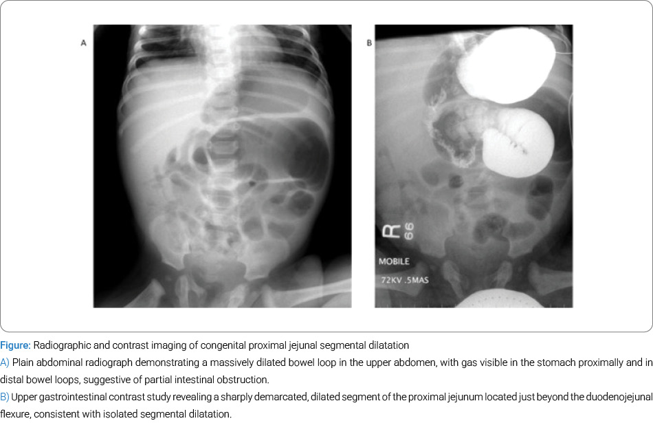
A one day old male neonate, born at term weighing 3600 g, was delivered spontaneously following antenatal findings of polyhydramnios and a dilated bowel loop suggestive of intestinal obstruction. Initial evaluation was performed at a district hospital before transfer to our Neonatal Intensive Care Unit (NICU). The infant developed bilious vomiting immediately after birth. He was clinically stable, with upper abdominal distension that was soft and non tender, and no palpable mass. Blood counts and biochemical profiles were normal. Post natal ultrasound confirmed a massively dilated proximal jejunal segment with no associated anomalies. Plain abdominal radiograph revealed a massively dilated bowel loop in the upper abdomen, with gas visible both proximally in the and distally in the pelvis (Figure A).
Differential diagnoses included acute intestinal obstruction, volvulus, Hirschsprung’s disease, and midgut malrotation. To exclude midgut volvulus, a potentially lethal condition, a water soluble contrast study was performed via nasogastric tube. An upper gastrointestinal contrast study confirmed a sharply demarcated dilated segment of the proximal jejunum just beyond the duodenojejunal flexure. This confirmed an adynamic, massively dilated, isolated proximal jejunal loop without mechanical obstruction, with abundant bowel gas distally in the abdomen and pelvis (Figure B).
The infant was resuscitated with intravenous fluids and nasogastric suction. Emergency exploratory laparotomy via a minimally invasive periumbilical incision revealed a hugely dilated isolated proximal jejunal segment distal to the duodenojejunal flexure. No obstructing lesion was present, and bowel contents could be moved freely.
Resection of the dilated segment followed by end to end jejunojejunostomy was performed uneventfully. Post operative recovery was smooth. Breastfeeding commenced on day 5, and discharge occurred on day 7. At 20 year follow‑up, the patient remains well and asymptomatic.
Discussion
Congenital segmental jejunal dilatation presenting as neonatal intestinal obstruction is rare. Congenital Segmental Intestinal Dilatation is characterized by a localized, sharply demarcated dilated segment of intestine without intrinsic or extrinsic obstruction, abnormal innervation, or transition zone.
In neonates, CSID may mimic midgut volvulus, Hirschsprung’s disease, or functional obstruction [3]. Jejunal involvement is uncommon, and its etiology remains unclear despite several proposed mechanisms [4]. These include intrauterine vascular accidents [5], associations with multiple intestinal atresias [6], and external compression of the fetal bowel [7].
Swenson and Rathauser (1959) [8] established diagnostic criteria:
• Abrupt transition between dilated and normal bowel.
• No intrinsic or extrinsic barrier distal to the dilatation.
• Limited bowel dilatation with a 3‑ to 4‑fold increase in size.
• Clinical presentation of occlusion or sub‑occlusion.
• Complete recovery after resection.
• Normal neuronal plexus.
A nationwide retrospective cohort study reported 28 cases of CSID [9,10]. The ileum was affected in 14 cases, the colon in 10, and the jejunum rarely. Surgical resection with end‑to‑end anastomosis was the treatment of choice in nearly all cases, with good prognosis.
Histological findings vary, including ectopic tissue and abnormal muscle layers. Rarely, immature ganglion cells have been reported in isolated jejunal dilatation [11]. Segmental stenosis with neonatal perforation has also been described [12]. Surgical resection remains the definitive treatment, with excellent outcomes when no associated anomalies are present.
Conclusion
This case highlights an unusual and rare presentation of isolated proximal jejunal segmental dilatation diagnosed prenatally and treated successfully in the immediate neonatal period. Resection and end to end anastomosis resulted in an excellent long term prognosis.
Acknowledgement
We are grateful to Mr. Clive W. Hardy for proofreading our article and making extensive language corrections. We are grateful to Miss Indre Zaparackaite, Consultant Pediatric Surgeon at the Evelina Children’s Hospital, London; Mr. Shailinder Singh, Consultant Pediatric Surgeon at the University of Nottingham NHS Foundation Trust, Nottingham, UK; Prof. C K Sinha, Consultant Pediatric Surgeon at St George’s Hospital, London; and Dr. Debashish Bhattacharya, Chief Pediatric Surgeon at Sultan Qaboos University Teaching Hospital, Salalah, Oman, who were involved in drafting, revising, and editing the manuscript critically for important intellectual content.
Conflict of Interests
The authors declare no potential conflicts of interest with respect to the research, authorship, and/or publication of this article. Informed consent was obtained for this publication.
References
- Shah AA, Shah AV. Congenital segmental dilatation of jejunum: a rare entity. J Indian Assoc Pediatr Surg. 2019;24(4):285–287.
- Patel RV. Congenital proximal jejunal diaphragm. J Indian Med Assoc. 1996;94(3):115–121.
- Hosie S, Lorenz C, Schaible T, Weber G, Waag KL. Segmental dilatation of the jejunum resembling prenatal volvulus. J Pediatr Surg. 2001;36(6):927–929.
- Kasanga TK, Zeng FT, Jampy‑Biaya S, Mbuyi AN, Mbuyi‑Musanzayi S. Congenital segmental dilatation of the jejunum in an African child: a case report. Pan Afr Med J. 2021;38:122.
- Patel RV, Trambadia RA, Anthony FM, Chhaniara RA, Govani D, Patel RR, et al. Case series—congenital neonatal colonic atresias arising in watershed areas of the colonic blood supply. Journal of the Indian Medical Association (JIMA). 2024;122(2):55–60.
- Harjai MM, Katiyar A, Negi V, Yadav D, Sharma M. Congenital segmental dilatation of jejunoileal region in a newborn: unusual clinical and radiologic presentation. J Indian Assoc Pediatr Surg. 2010;15(3):96–97.
- Patel RV, Deshmukh SS, Gandhi RK, Kadam NN, Kadam SN, Arora HL. Multiple ileal atresia: a unique technique of prograde and retrograde midgut decompression. J Indian Assoc Pediatr Surg. 2000;5(2):58–61.
- Swenson O, Rathauser F. Segmental dilatation of the colon; a new entity. Am J Surg. 1959;97(6):734–738.
- Sergi C, Hager T, Hager J. Congenital Segmental Intestinal Dilatation: A 25-Year Review with Long-Term Follow-up at the Medical University of Innsbruck, Austria. AJP Rep. 2019;9(3):e218–e225.
- Sakaguchi T, Hamada Y, Masumoto K, Taguchi T; Japanese Study Group of Allied Disorders of Hirschsprung’s Disease. Segmental dilatation of the intestine: results of a nationwide survey in Japan. Pediatr Surg Int. 2015;31(11):1073–1076.
- Adhikari SB, Rana S, Chalise S. Diagnostic dilemma of congenital segmental jejunal pseudo‑dilatation associated with immature ganglion cells in the colon: a case report. J Neonatal Surg. 2020;9:13.
- Lawther S, Patel RV, de la Hunt MN. Ileal duplication cyst associated with segmental ileal stenosis and neonatal perforation. J Pediatr Surg Case Rep. 2013;1(1):8–10.
Cite this article
Govani DR, Singh H, Swamy KB, Midha PK, Govani ND, Panchasara NG, et al. Congenital Neonatal Proximal Jejunal Isolated Segmental Dilatation. Clin Surg J. 2026;7(2):1–3.
Keywords
Acute abdomen; Neonatal; Prenatal; Congenital; Intestinal obstruction; Jejunal; Segmental dilatation; Periumbilical laparotomy
Copyright
© 2026 Ramnik Patel. This is an open access article distributed under the terms of the Creative Commons Attribution 4.0 International License (CC BY-4.0).