Abstract
Cancers of the appendix are very rare, and among them, Signet Ring Cell Carcinoma of the Appendix (SRCCA) is the rarest type, with an incidence of about 4%. SRCCA was first described in 1969. The most common age at diagnosis is 62 years, with a 1:11 ratio indicating a female predominance. The etiology and risk factors for epithelial tumors of the appendix are not clearly known. The tumor cells contain a large vacuole that pushes the nucleus to the periphery, giving them a typical signet-ring appearance. According to the WHO, an appendiceal tumor with more than 50% signet-ring cells is classified as SRCCA. The clinical features of SRCCA are nonspecific, and there are likewise no specific radiological findings for its diagnosis. Treatment may include a simple appendectomy or a right hemicolectomy, depending on tumor invasion. The role of adjuvant chemotherapy and radiotherapy remains debatable. Most major centers prefer a combination of cytoreductive surgery with hyperthermic intraperitoneal chemotherapy.
Abbreviations
SRCCA: Signet Ring Cell Carcinoma of the Appendix; GI: Gastointestinal; HPE: Histopathology Examination; IHC: Immunohistochemistry; RH: Right Hemicolectomy; Lymph Node Dissection
Introduction
Cancers of the appendix are uncommon, accounting for only about 0.4% of all Gastrointestinal (GI) malignancies [1,2]. Appendiceal malignancies can be broadly divided into neuroendocrine neoplasms (50%–75%), previously called carcinoid tumors [3,4], and adenocarcinomas. Appendiceal adenocarcinomas are further classified into three groups: mucinous adenocarcinoma, low-grade appendiceal mucinous neoplasms, and signet ring cell carcinomas [4]. Among these, signet ring cell carcinoma of the appendix (SRCCA) is an exceedingly rare entity [1]. SRCCA was first described in 1969 [3], and the World Health Organization classifies it as an adenocarcinoma composed of more than 50% isolated malignant cells containing intracytoplasmic mucin [1]. We present a case of SRCCA that manifested as an appendiceal mucocele.
Case Presentation
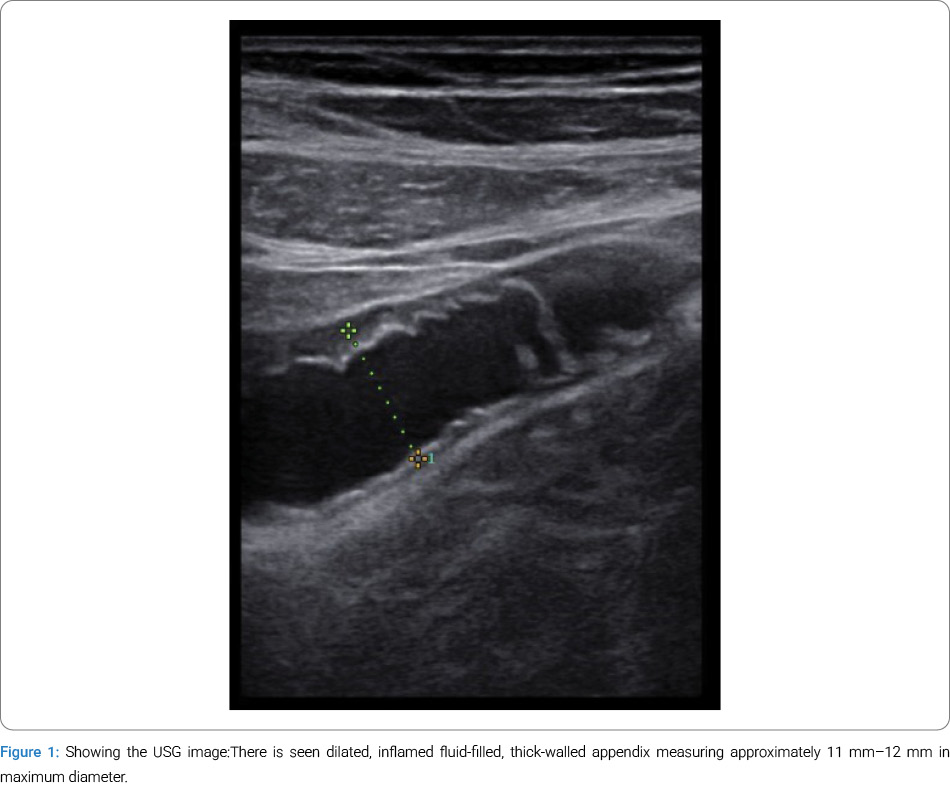
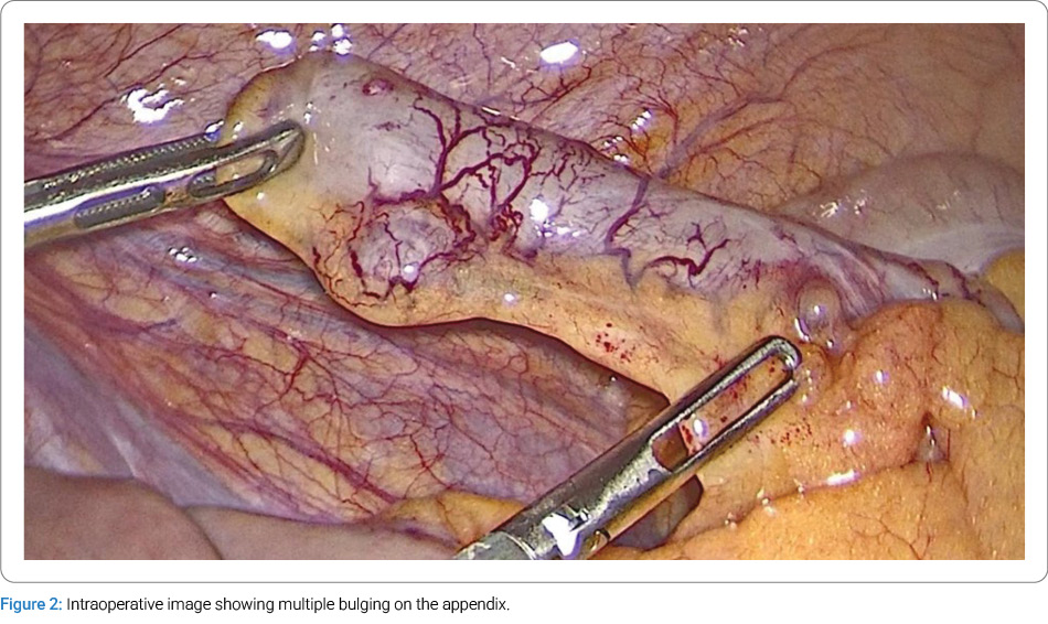
A 54-year-old man presented to the emergency department with a two-day history of right lower abdominal pain. The pain began suddenly, without any history of trauma. It was continuous and progressively worsening, and was associated with nausea. There were no aggravating or relieving factors. He was a known diabetic on regular medication. On examination, the patient was afebrile and not tachycardic. Abdominal examination revealed tenderness at McBurney’s point and positive rebound tenderness. No palpable mass was detected in the right iliac fossa. Ultrasonography of the abdomen revealed a dilated, inflamed, fluid-filled, thick-walled appendix measuring 11 mm–12 mm in maximum diameter, with periappendiceal fat stranding in the right iliac fossa, suggestive of acute appendicitis with a developing mucocele (Figure 1). Based on this diagnosis, we proceeded with a laparoscopic appendectomy. Intraoperatively, we noted multiple bulges on the appendix (Figure 2), prompting us to consider appendiceal diverticulosis as one of the differential diagnoses, though it is rare. No other significant findings were noted in the intestines, peritoneal surfaces, pelvic cavity, or on the liver surface, and the surgery was uneventful.
The postoperative course was also uneventful, and the patient was discharged the next day due to health insurance requirements. The HPE report indicated that the appendiceal wall contained pools of mucin with scattered signet ring cells. The tumor is seen infiltrating the muscularis propria and subserosal fat. Perineural invasion has been identified. There is no evidence of lymphovascular invasion. The base of the appendix shows no evidence of dysplasia or malignancy. Due to limited resources, we were unable to perform an Immunohistochemistry (IHC) study. We planned a metastatic workup and a Secondary Right Hemicolectomy with Lymph Node Dissection (RH-LND). Still, as the patient is an expatriate, he returned to his home country for further treatment.
Discussion
According to the National Cancer Institute’s Surveillance Epidemiology data, SRCCA is the rarest variety, with a 4% incidence [4]. The most common age at diagnosis is 62 years, with a 1:11 ratio showing female predominance [5]. The etiology and risk factors for epithelial tumors of the appendix are not clearly known [6]. SRCCA is thought to arise from a mutation in pluripotent intestinal crypt epithelial cells, resulting in the formation of mucin droplets and neuroendocrine secretory granules within the epithelial intestinal crypt. The origin of SRCCA is primarily sporadic, but it is occasionally associated with conditions such as hereditary nonpolyposis colorectal cancer (Lynch syndrome). The tumor cells contain a large vacuole that pushes the nucleus to the periphery, giving them a characteristic signet-ring appearance [5]. According to the WHO, appendiceal tumors with more than 50% signet-ring cells are classified as SRCCA [4,5,7,8]. Abushalha et al. reported that most patients in their study were white and found no statistically significant differences in survival based on race [1].
The clinical features of SRCCA are nonspecific and often mimic acute appendicitis (most commonly), ovarian torsion, or other right lower abdominal pathologies [3]. Other nonspecific symptoms include weight loss, changes in bowel habits, or manifestations related to metastatic disease [5]. Signet Ring Cell Carcinoma (SRCC) most commonly affects the stomach [4]. Due to the proximity of the terminal ileum, ileocecal valve, cecum, appendix, and lymphatic connections, the pathology may involve multiple sites, resulting in delayed diagnosis. Lymphatic metastasis can also mimic inflammatory conditions, adding further uncertainty. In IHC, CDX-2 and CK20 help distinguish SRCCA from other SRCCs [4,5].
Radiologically, CT or MRI can aid in detecting malignancy, but findings often resemble acute appendicitis [5]. There are no characteristic features of SRCCA; therefore, diagnosing or differentiating it from other tumor types is practically impossible. A mucinous epithelial tumor of the appendix may appear as a mucocele on CT. In fewer than 50% of cases, linear mural calcification is present, along with less common features such as soft-tissue thickening, irregularity of the mucocele wall, and adjacent lymph nodes [9].
Terada reported a case in which the patient was initially diagnosed with acute appendicitis based on clinical, laboratory, and radiologic findings. On histopathology, SRCCA was identified in the proximal appendix, while the remainder of the appendix showed features of acute phlegmonous appendicitis. The author suggested that narrowing of the lumen caused by the tumor may have resulted in acute appendicitis.
SRCCA is considered a highly aggressive variant and is frequently diagnosed at a metastatic stage. In 93% of cases, it metastasizes to adjacent organs, lymph nodes, or the peritoneal cavity [9]. It carries a poor 5-year survival rate of 7% [4]. Its extremely low incidence and frequent presentation with metastasis contribute to its poor prognosis [2]. The two types of appendiceal tumors with the most significant impact on survival are SRCCA and malignant carcinoid [10].
The optimal surgical management of SRCCA remains unclear. Options to consider include:
a) Simple appendectomy: for SRCCA limited to the mucosa (identified postoperatively).
b) Right hemicolectomy with lymph node dissection (RH-LND): if SRCCA is diagnosed preoperatively (rare) or postoperatively.
There is no statistically significant difference in 5-year survival between localized and extended resection for mucosa-limited lesions; however, the opposite is true for lesions extending beyond the mucosa [5]. McGory et al. recommended an aggressive approach, such as RH for tumors larger than 2 cm, due to significantly better 5-year survival compared to tumors 1 cm or smaller [11]. Abushalha et al. observed similar results in their study. Still, multivariate analysis could not confirm these findings because tumor size was not documented in many case reports, necessitating its exclusion from the analysis.
The role of adjuvant chemotherapy and radiotherapy remains debatable [1]. According to the National Comprehensive Cancer Network (NCCN) guidelines, 5-fluorouracil–based chemotherapy should be considered for appendiceal malignancy, although its role in SRCCA is uncertain [5]. Anderson Cancer Center recommends systemic chemotherapy for metastatic SRCCA [1]. Following cytoreduction, adjuvant chemotherapy (The Capecitabine and Oxaliplatin regimen) has been shown to improve survival [2]. Treatment options for metastatic SRCCA include systemic chemotherapy alone, Hyperthermic Intraperitoneal Chemotherapy (HIPEC), cytoreductive surgery with peritonectomy, or combinations of these approaches. Most major centers prefer combining cytoreductive surgery with HIPEC [7]. Ko et al. suggested routine oophorectomy for all female patients, especially postmenopausal women. Due to the rarity of the disease, conclusive data regarding surgical debulking in diffuse metastatic cases are limited. For aggressive appendiceal malignancies, simple RH alone is not recommended; it should be combined with cytoreduction and intraperitoneal chemotherapy [10].
In conclusion, due to the rarity of SRCCA, it is very likely to be missed preoperatively. More studies are needed to establish a workup or treatment protocol. Still, until then, SRCCA should be considered a differential diagnosis in all elderly patients presenting with acute appendicitis or a mucocele. Even the slightest suspicion should prompt a full diagnostic workup.
Acknowledgement
We are thankful to the patient for allowing us to publish his case.
Conflict of Interest
The authors declare no potential conflicts of interest with respect to the research, authorship, and/or publication of this article. Informed consent was obtained for this publication.
Cite this article
Shyam DC. Signet ring cell carcinoma of the Appendix Duped as a Mucocele of the Appendix: A Case Report. Clin Surg J. 2026;7(2):1–4.
Keywords
Appendiceal tumor; Signet ring cell carcinoma of appendix; Secondary right hemicolectomy; Intraperitoneal chemotherapy
Copyright
© 2026 Devajit Chowlek Shyam. This is an open access article distributed under the terms of the Creative Commons Attribution 4.0 International License (CC BY-4.0).