Disadvantages of V-Y Flaps in Pressure Sores Reconstruction in Spinal Cord Injured Persons: A Case Series
* Luca Negosanti;
Laura Simoncini;
Siriana Landi;
Manuela Marani;
Micaela Battilana;
Rossella Sgarzani;
-
* Luca Negosanti: Montecatone Rehabilitation Institute, Imola, Italy
-
Laura Simoncini: Montecatone Rehabilitation Institute, Imola, Italy
-
Siriana Landi: Montecatone Rehabilitation Institute, Imola, Italy
-
Manuela Marani: Montecatone Rehabilitation Institute, Imola, Italy
-
Micaela Battilana: Montecatone Rehabilitation Institute, Imola, Italy
-
Rossella Sgarzani: Department of DIMEC, University of Bologna, Bologna, Italy
Abstract
Pressure sores are a frequent complication in spinal cord injury/ disease patients. Treatment usually consists of wide debridement of the wound, followed by reconstruction with local flaps. Various techniques have been described to repair these defects, including local fasciocutaneous flaps, muscle or myocutaneous flaps, and free flaps. The V-Y flap is a common reconstructive technique in plastic surgery and consists of the local advancement of an island flap with random vascularization from its subcutaneous pedicle. This flap was originally described for finger reconstruction, but its application has since been extended to numerous anatomical sites, including the face and perineal region, and it is frequently reported in pressure-sore reconstruction. In this report, we aim to highlight the potential disadvantages of using the V-Y flap in the reconstruction of pressure sores in patients with spinal cord injury.
Introduction
Pressure Sores (PS) are a frequent complication in patients with Spinal Cord Injury/Disease (SCI/D), with an incidence of 34% in individuals with quadriplegia and 47% in those with paraplegia [1]. In stage III and IV lesions, according to the National Pressure Ulcer Advisory Panel (NPUAP) classification [2], surgery is the main treatment [3].
Treatment usually consists of wide wound debridement followed by reconstruction with local flaps. Several reconstructive techniques have been described for the management of these defects [3–7]. The V-Y flap is a commonly used reconstructive technique in plastic surgery and consists of the local advancement of an island flap with random vascularization based on its subcutaneous pedicle [8–11]. Based on our clinical experience, this flap may present several limitations when used for PS reconstruction in SCI/D patients. In this report, we aim to highlight the potential disadvantages of the V-Y flap in PS reconstruction for SCI/D patients by analyzing possible risk factors for complications and conceptual limitations associated with its use in this specific patient population.
Case Presentation
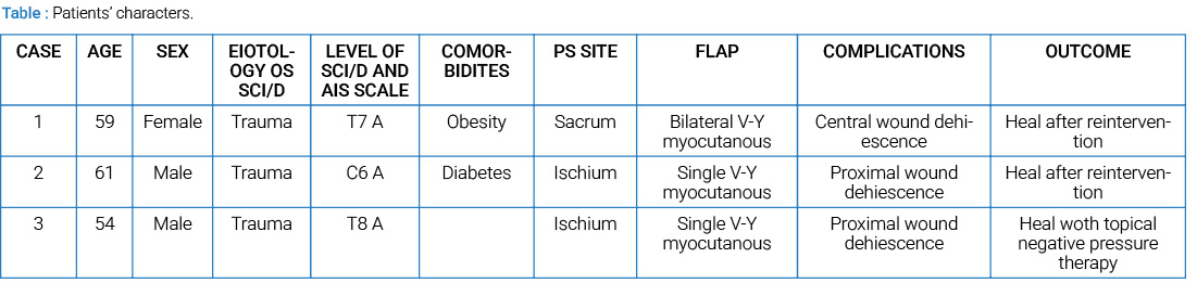
We report three cases of SCI/D patients affected by PS and treated with V-Y flaps (Table).
Case 1
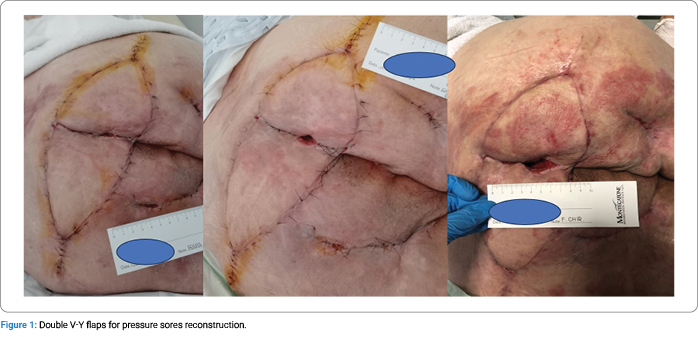
A 59-year-old female patient affected by post-traumatic paraplegia at level T7, Abbreviated Injury Scale (AIS) A [12], was treated at another hospital for a sacral PS and reconstructed using bilateral V-Y myocutaneous flaps. Immediately after surgery, wound dehiscence occurred in the central portion of the surgical site, corresponding to the area requiring the greatest reconstructive coverage. After surgical debridement, topical negative pressure therapy was applied, resulting in a reduction of the dehiscence (Figure 1). A secondary surgical procedure was subsequently planned.
Case 2
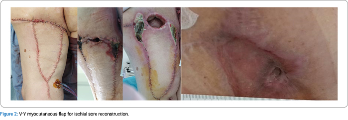
A 61-year-old male patient affected by post-traumatic quadriplegia at level C6, AIS A [12], was treated at another center for an ischial PS and reconstructed with a V-Y myocutaneous flap. He was transferred to our hospital 10 days after surgery. Partial flap necrosis occurred, with extensive dehiscence interested therapyall the entire proximal portion of the flap. Immediate surgical debridement was performed, followed by application of topical negative-pressure wound therapy. Fortunately, the remaining viable muscle tissue was sufficient to cover the bone prominence, and a split-thickness skin graft was subsequently harvested and applied (Figure 2).
Case 3
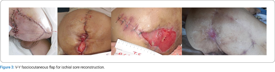
A 54-year-old male patient, affected by post-traumatic paraplegia at level T8, AIS A, was treated at our hospital for the third recurrence of an ischial PS. The wound was surgically debrided and reconstructed using a V-Y myocutaneous flap. Two weeks after surgery, wound dehiscence was observed at the proximal portion of the flap, involving the skin and subcutaneous tissue. Fortunately, the residual viable muscle tissue adequately covered the bone. Topical negative-pressure wound therapy was applied, resulting in a reduction in wound size (Figure 3). The patient was discharged with instructions to continue wound care treatment, and the wound healed by secondary intention.
All patients were treated in accordance with our internal postoperative care protocol for PS reconstruction, as previously reported [6].
Discussion
The use of the V-Y random advancement flap is frequently described in reconstruction following PS debridement. However, the reported cases suggest that this technique may not be sufficiently safe for SCI/D patients. The main limitation of this study is the small number of reported patients; nevertheless, several considerations can be drawn. The first factor to consider is spasticity, which is present in more than 60% of SCI/D patients [13–15]. Muscle spasms may cause detachment of the proximal portion of the flap along different tissue planes, resulting in deep wound dehiscence. Therefore, spasticity must be considered, and flap design should aim to minimize tension during spasms [16]. Another relevant aspect is vascular dysfunction following SCI/D [17]. This condition may lead to delayed hematoma formation, flap detachment, and fistula development. The vascular supply of flaps in SCI/D patients may differ from that in other patient populations due to chronic hypotension. Disruption of descendent autonomic pathways in SCI/D results in sympathetic hypoactivity, causing hypotension and impaired regulation of blood pressure variability. These factors increase the risk of reduced vascular supply to the flap. Based on our experience, it is preferable to harvest the shorter flap necessary to reduce the risk of distal flap necrosis. Additionally, longer scars, such as those resulting from a V-Y flap, may pose challenges in the event of recurrence by limiting the availability of alternative reconstructive options. Bilateral V-Y myocutaneous flaps for sacral PS compromise both donor sites;therefore, in the event of flap failure or recurrence, reconstruction becomes particularly challenging. For this reason, a unilateral fasciocutaneous flap should be considered the first-line option for sacral PS reconstruction [18]. In the series reported by Nööjd et al. on 111 flaps used for PS reconstructions, a 90% survival rate was reported, and they conclude that the V-Y flap is preferable to the axial flap [19]. In this series, 29 patients without SCI/D were included, and the reported recurrence rate was also high (16% at 1 year). In our experience, we prefer local transpositional fasciocutaneous flaps for PS reconstruction. We have previously reported our experience with favorable outcomes in terms of complication rate [6,18]. A transpositional flap provides adequate defect coverage and can be designed with postoperative bed positioning and associated spasticity in mind, thereby reducing the risk of wound dehiscence. In addition, residual scars resulting from transpositional flaps are less extensive than those created by the V-Y flap, allowing for greater reconstructive options in the event of recurrence.
The main limitation of this report is the small number of patients. Nevertheless, we believe that these cases provide meaningful insights that allow specific considerations related to SCI/D patients undergoing PS reconstruction. SCI/D patients present unique characteristics that must be considered during reconstructive planning to reduce the complication rate. Larger series are needed to accurately define the true risk associated with the V-Y flap in PS reconstruction in SCI/D and to identify specific risk factors to optimize reconstructive planning.
Conclusion
Patients with SCI/D represent a particularly complex population, and multiple factors must be considered by reconstructive surgeons when treating PS. Spasticity, vascular dysfunction, post-operative bed positioning, and the high risk of recurrence are among the most important aspects to be addressed during reconstructive planning to reduce the complication rate. V-Y flaps appear to be a suboptimal option in this patient population unless carefully selected and meticulously planned. Further studies involving larger patient series are required to better understand postoperative complications and to identify reconstructive strategies.
Acknowledgements
The authors thanks Sara Nesti for language revision.
Conflicts of Interest
The authors declare no potential conflicts of interest with respect to the research, authorship, and/or publication of this article. Informed consent was obtained for this publication.
References
- Cowan LJ, Ahn H, Flores M, Yarrow J, Barks LS, Garvan C, et al. Pressure ulcer prevalence by level of paralysis in patients with spinal cord injury in long-term care. Adv Ski Wound Care. 2019;32(3):122–130.
- Edsberg LE, Black JM, Goldberg M, McNichol L, Moore L, Sieggreen M, et al. Revised national pressure ulcer advisory panel pressure injury staging system: revised pressure injury staging system. J Wound Ostomy Cont Nurs. 2016;43(6):585–597.
- Chen CY, Chiang IH, Ou KL, Chiu YL, Liu HH, Chang CK, et al. Surgical treatment and strategy in patients with pressure sores: A single-surgeon experience. Medicine (Baltimore). 2020;99(44):e23022.
- Oksman D, de Almeida OM, de Arruda RG, de Almeida MLM, do Carmo FS. Comparative study between fasciocutaneous and myocutaneous flaps in the surgical treatment of pressure ulcers of the sacral region. JPRAS Open. 2018;16:50–60.
- Negosanti L, Tedeschi S, Trapani FF, Gaiani L, Sgarzani R. Multiple flaps for trochanteric pressure sore reconstruction: a case series. Cureus. 2020;12(7):e9369.
- Sgarzani R, Maietti E, Tedeschi S, Trapani FF, Battilana M, Landi S, et al. Multidisciplinary treatment protocol for ischiatic, sacral, trochanteric or other pressure injuries in people with spinal cord injury: a retrospective cohort study. Spinal Cord. 2023;61(3):204–210.
- Lemaire V, Boulanger K, Heymans O. Free flaps for pressure sore coverage. Ann Plast Surg. 2008;60(6):631–634.
- Tranquilli-Leali E. Ricostruzione dell’apice delle falangi ungueali mediante autoplastica volare peduncolata per scorrimento. InfortTraumatol Lav. 1935;1:186–193.
- Pribaz JJ, Chester CH, Barrall DT. The extended V-Y flap. Plast Reconstr Surg. 1992;90(2):275–280.
- Giannini A, Di Donato V, D’Oria O, Schiavi MC, May J, Benedetti Panici P, et al. The V-Y gluteal fold advancement flap: Outcomes following radical surgery for vulvar malignancies. Int J Gynaecol Obstet. 2021;152(3):421-424.
- Djedovic G, Metzler J, Morandi EM, Wachter T, Kühn S, Pierer G, et al. Comparison of fasciocutaneous V-Y and rotational flaps for defect coverage of sacral pressure sores: a critical single-centre appraisal. Int Wound J. 2017;14(6):945–949.
- American Spinal Injury Association. ASIA Impairment Scale [Internet]. Richmond (VA): The American Spinal Injury Association; 2026.
- Levi R, Hultling C, Seiger A. The stockholm spinal cord injury study: 2. Associations between clinical patient characteristics and post-acute medical problems. Paraplegia. 1995;33(10):585–594.
- Lance J. Pathophysiology of spasticity and clinical experience with baclofen. In: Lance J, Feldman R, Young R, Koella W, editors. Spasticity: Disordered Motor Control. Chicago(IL): Yearbook;1980. p. 185–204.
- Little J, Micklesen P, Umlauf R, Britell C. Lower extremity manifestations of spasticity in chronic spinal cord injury. Am J Phys Med Rehabil. 1989;68(1):32–36.
- Israel JS, Carlson AR, Bonneau LA, Kempton SJ, King TW, Bentz ML, et al. Reconstructive surgery and patients with spinal cord injury: Perioperative considerations for the plastic surgeon. J Plast Surg Hand Surg. 2016;50(1):44–49.
- Popa C, Popa F, Grigorean VT, Onose G, Sandu AM, Popescu M, et al. Vascular dysfunctions following spinal cord injury. J Med Life. 2010;3(3):275–285.
- Sgarzani R, Rucci P, Landi S, Battilana M, Capirossi R, Aramini B, et al. Reconstructive surgery of pressure injuries in spinal cord injury/disorder patients: retrospective observational study and proposal of an algorithm for the flap choice. Healthcare (Basel). 2023;12(1):34.
- Nööjd M, Wyckman A, Steinvall I, Elmasry M. Flap survival after reconstructive surgery for pressure ulcers: a cohort study. Plast Reconstr Surg Glob Open. 2023;11(12):e5451.
Cite this article
Negosanti L, Simoncini L, Landi S, Marani M, Battilana M, Sgarzani R. Disadvantages of V-Y flaps in pressure sores reconstruction in spinal cord injured persons: a case series. Clin Surg J. 2026;7(2):1–4.
Keywords
V-Y flap; Pressure sores; Spinal cord injury; Spasticity; Reconstructive surgery; Wound care; Local flaps
Copyright
© 2026 Luca Negosanti. This is an open access article distributed under the terms of the Creative Commons Attribution 4.0 International License (CC BY-4.0).